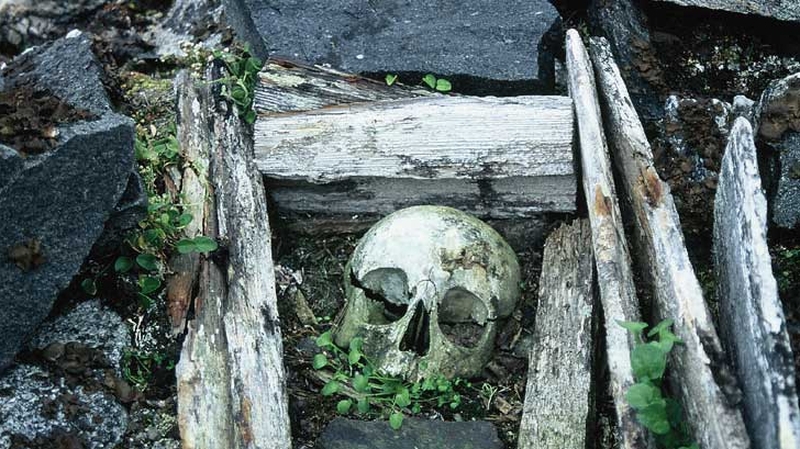
The remains of a 17th century whaler a roughly-hewn wooden coffin on the western coast of Spitsbergen, in the Arctic's Svalbard Archipelago. The burial has resurfaced due to active periglacial processes (Pic: Vincent Butler)
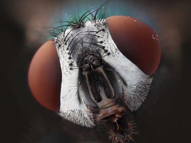
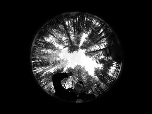
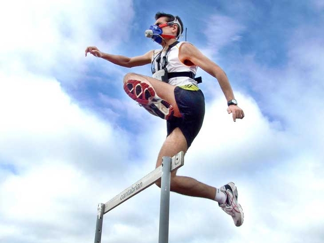
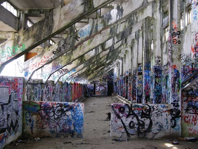
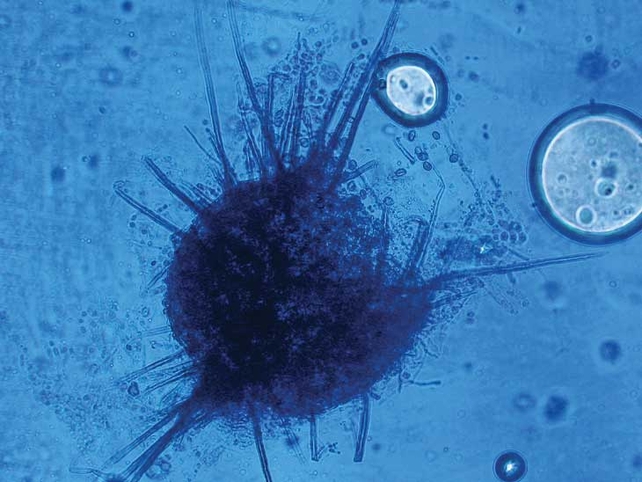
A common green bottle fly shot on a self-constructed optical microscope, made as as an affordable alternative to similar more expensive microscopes (Pic: Karl Gaff)A mature Sitka spruce stand at Mount Callan, Co Clare, captured using a technique called hemispherical photography, also known as fisheye photography (Pic: Denis Coghlan)A photo taken during testing to understand the body’s response to exercise in the real world (Pic: Ciara O’Hagan)This is an image of a graffiti gallery prototype in the baths in Blackrock Co Dublin. It was captured during research into the movement of graffiti as vandalism, to graffiti as accepted works of art (Pic: Connell Vaughan)A fern spore analysed by Scanning Electron microscopy. The colour has
been modified (Pic: Dr Alfonso Blanco)
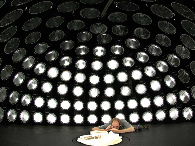
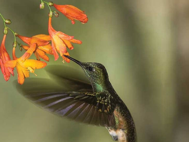
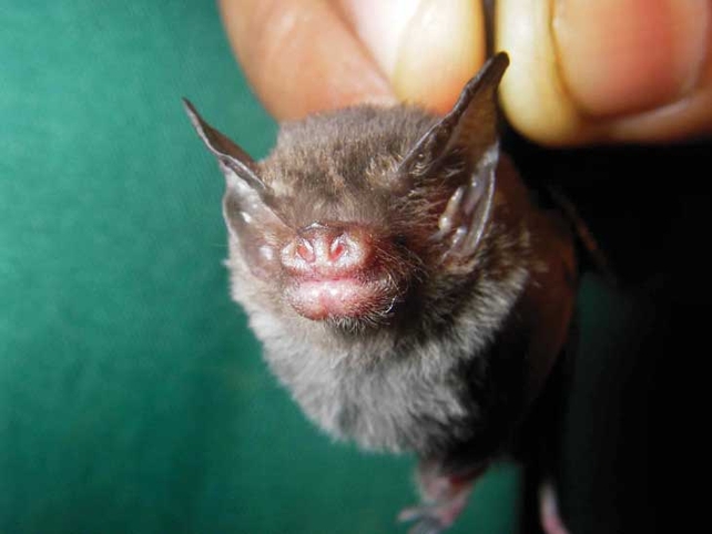
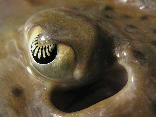
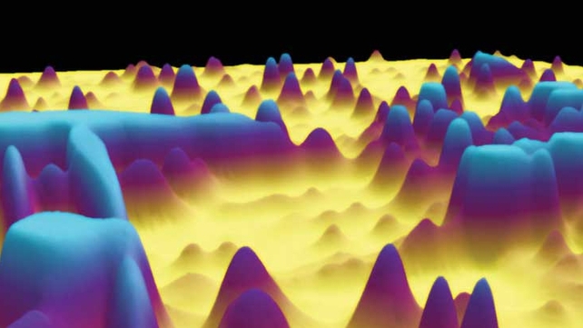
This photo was taken of a researcher under Ireland’s only artificial sky. He is studying the internal visual environment in an architectural model under 145 individually controlled lamps (Pic: Paul Kenny)A hummingbird hovering at a flower, captured during research into the study of bird flight and its evolution (Pic: Billy Clarke)Fungi Chaetomium speceimen isolated from an eye infection in a horse. It has been stained cotton blue with lactophenol so that a microbiologist can clearly see it under the microscope. (Pic: Yvonne Abbott)This is a photo of the Craseonycteris thonglongyai (bumble-bee bat). It is one of the rarest species in the world and is only found in two populations, one in Thailand and one in Myanmar (Pic: Emma Teeling)The eye of a spotted ray (Raja montagui) caught during a survey of the Irish Sea aboard the RV Celtic Voyager. Its intricately shaped iris makes the ray’s eye very sensitive to movements within a large visual field (Pic: Edward Farrell)An image of a protein extract from a breast cancer cell line. Protein cells have been separated on a gel and scanned. Each individual protein appears as a peak where height represents its abundance (Pic: Thomas Lau)